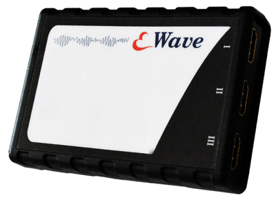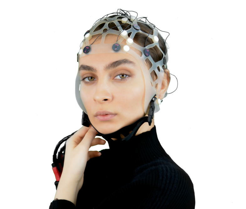
eWave+ is a multi-channel bio-signal amplifier, which has 32, 40, 64, or 128 recording channels. eWave+ provides a wide input sensitivity to record various bio-signals such as EEG, EMG, ECG, and EOG. Besides, eWave+ is a special ERP acquisition system with high precision. Sciencebeam has also offered eCap series of EEG caps to provide consistent signal recording of up to 128 electrodes. Furthermore, external body sensors can be connected in order to record various biological signals. eProbe software, which is a user-friendly software that is compatible with eWave+ device, is also offered by Sciencebeam company for visualization and analysis of recorded signals. Also, all 128 channels can be analyzed in real-time with eBridge Simulink software.
HOSPITAL EEG
EEG, which is a non-invasive method for measuring brain electrical activity, helps clinicians in diagnosing conditions affecting brains such as head injuries, headaches, brain tumors, sleep problems and etc. The eWave+ device can be used in hospitals so as to help clinicians diagnose special conditions. Besides, due to eProbe software’s abilities, it is possible to import the recorded data into MATLAB, EEGLAB, and LABVIEW environments for processing purposes.
SOFTWARE AND PLUGINS
eWave+ is compatible with a wide range of software in order to meet the needs of various users including engineers, researchers, scientists, clinicians, and psychologists. Include MATLAB, LABVIEW, NeuroGuide, Tobi, etc…
Size: 120 (L) × 120 (W) × 41 (H)
Weight: 318 gr
Interface: USB, Wifi
Digital inputs: 4 digital trigger inputs
Supply: 5V DC, Lithium battery
Bandwidth: 200 Hz
Resolution: 24-bit
Noise level: less than 0.5 μνrms
Amplifier type: DC, differential
Input impedance: 109 Ω
Safety class: ǁ
Standards: IEC, CE, ISO13485, ISO9001
Transcranial Magnetic Stimulation (TMS), which is a non-invasive form of brain stimulation, uses a changing electromagnetic field to cause electric current at a specific area of the brain. Due to the latest developments, simultaneous recording of EEG and TMS has become possible. The combination of TMS with simultaneous EEG provides the possibility to non-invasively probe the brain’s excitability in real-time.
Transcranial Direct Current Stimulation (tDCS) is a form of brain stimulation, where very low levels of constant current are delivered to specific areas of the brain. Basically, tDCS is developed to help patients with brain injuries or psychological conditions such as depression. The combination of tDCS with simultaneous EEG provides valuable information about tDCS mechanisms. The combined EEG-tDCS system can also be used for the preventive treatment of neurological conditions. eWave+ device has provided a simultaneous recording of EEG with TMS and tDCS.


ERP (Event-Related Potentials) are small Changes in scalp recorded EEG that are time-locked to an onset Auditory or visual stimulus. ERP is used to investigate the process of information in the brain.
In ERP recording results, there is a time gap between stimulus presentation and the point of “Max” value (peak). Presumable components in the laptop that the time-gap or delay occurs are; CPU, motherboard, and graphic card. This delay depicts the time taken by the stimulus information to generate the ERP results. To prevent this pause and get a precise result in a recording; We have designed a module in our eWave+ that omits the delay and enables the ERPs recording to happens in realtime and leaves no gap between stimulus presentation and the subject’s experience.
Other ERP acquisition systems record ERP as command executes from the monitor, except eWave+, which records stimulus presentation synchronously in ERP Results. This exclusive feature only exists in eWave+ and makes it the most explicit ERP acquisition system in the world.
A BCI system is a communication channel between a brain and a computer. This computer-based system acquires brain signals, analyzes them, and converts them into actions including hand or leg movement, opening or closing a door, and other daily activities. A BCI system especially improves the quality of life of disabled patients and makes it possible for them to interact with their environment. Connecting Smart Box to eWave+ provides an efficient BCI system that is MATLAB-based and includes all required parts for data recording, real-time analysis, and also data classification. Also, you can import data in Simulink directly by using eBridge environment, where visualization, feature extraction, and classification of your data are possible by using the Simulink blocks.

High-Frequency Oscillations (HFOs) usually occur in epilepsy patients which can be recorded with the Electrocorticogram (EcOG) with the implemented electrode grids in their brains. Recording HFOs requires a high-performance bio-signal amplifier with high sampling frequency and a very good signal-to-noise ratio (SNR) and resolution. Mapping neural networks are important for pre-surgical planning in epilepsy patients. Since HFOs can be found in such neural networks, brain surgeons need to distinguish physiological HFOs from pathological ones.
Due to eWave+’s high sampling frequency (1000 Hz) and very good SNR, it has the ability to record HFOs. ECoG electrodes from Ad-Tech, PMT, Unique Medical, and Cortec can be used with eWave+.

Sleep is generally characterized by a reduction involuntary body movement decreased to a little reaction to external stimuli, loss of consciousness, a reduction in auditory receptivity, an increased rate of anabolism (the synthesis of cell structures), and a decreased rate of catabolism (the breakdown of cell structures). The capability for arousal from sleep is a protective mechanism and also necessary for health and survival.
Sleep progresses throughout the night in cycles of REM and NREM phases. In humans, these cycles are approximately 90 to 120 minutes long and each phase may have a distinct physiological function. Drugs such as alcohol and sleeping pills can suppress certain stages of sleep (see Sleep deprivation). This can result in a sleep that exhibits loss of consciousness but does not fulfill its physiological functions.
In REM sleep, the brain is active and the body inactive, and this is when most dreaming episodes occur. REM sleep is characterized by electroencephalography (EEG) that has low voltage and mixed frequencies, similar in appearance to the awake EEG. During REM sleep the sympathetic nervous system is active, but there is a loss of skeletal muscle tone and our muscles are paralyzed so that we don’t act out our dreams.
In NREM sleep, the body is active, while the brain is relatively inactive compared to REM sleep, and there is relatively little dreaming. Non-REM encompasses four stages; stages 1 and 2 are considered ‚light sleep‘, and 3 and 4 ‚deep sleep‘. They are differentiated solely using EEG and unlike during REM sleep which is characterized by rapid eye movements and a relative absence of muscle tone, during NREM sleep limb movements are quite frequent, and sleepwalking (parasomnia) can occur in non-REM sleep.
NEED MORE INFORMATION ABOUT THIS PRODUCT?
Send us your email so we can contact you as soon as possible.
Click one of our contacts below to chat on WhatsApp